Our lab has a long-standing interest in tumor initiation and progression. A better understanding on how tumors evolve over time with respect to genetic, epigenetic, and gene expression changes will elucidate the molecular mechanisms underlying the aggressiveness and fatality of the disease and provide insights on tumor initiation, metastasis, and drug resistance. Our research focuses on the two major subtypes of lung cancer, namely non-small cell lung cancer (lung adenocarcinoma) and small cell lung cancer. In the past we have identified and characterized protein-coding and non-coding (e.g. microRNAs) gene regulation in tumor progression, invasion and metastasis and this includes (1) loss of the master pulmonary differentiation factor Nkx2-1, (2) gain of the embryonic proto-oncogene Hmga2, (3) isoform switching of the invadopodia component Tks5, and (4) misregulation of the miR-200 family of microRNAs.
We have started to Investigate how maintaining protein homeostasis affects tumor evolution and progression. Cancer cells are continuously exposed to protein folding stress due to increased biosynthetic demands, hyperproliferation, mutational burden, immune attack, and oxidative stress. To combat these stressors, cancer cells exhibit elevated activity of protein homeostasis mechanisms to maintain the proteome in a folded and functional state. We are investigating how two critical mediators of protein homeostasis, Heat shock protein 90 (Hsp90), and Heat shock factor 1 (Hsf1) enable lung tumors to evolve and adapt while encountering diverse cellular stresses that impact protein folding. These studies will uncover unique characteristics of protein folding mechanisms in cancer cells that may be exploited for pharmacological intervention.

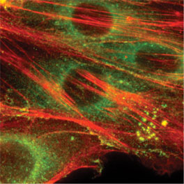
Invadopodia are proteolytic cellular protrusions that can degrade various components of extracellular matrix and are thought to enable tumor cells to metastasize. Shown here is an immunofluorescence image of metastatic lung adenocarcinoma cells with abundant invadopodia. The foci of invadopodia contain F-actin (red) and cortactin (green).

